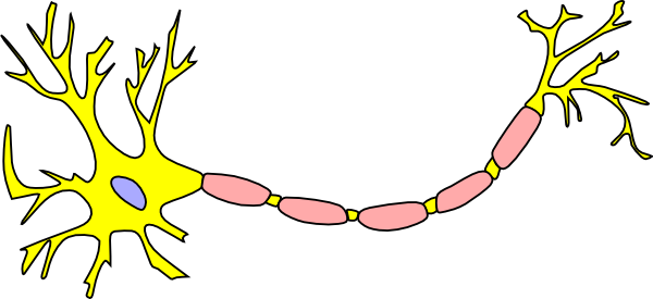
45 picture of a neuron without labels
diagram of eye with labels Neuron B&w Clip Art At Clker.com - Vector Clip Art Online, Royalty Free neuron clipart diagram nerve unlabeled clip cell neurons blank system digestive axon vector unlabelled motor cliparts 20clipart clipground clker royalty 32 Eye Diagram To Label - Labels Database 2020 ardozseven.blogspot.com Yvonnes neuropsychology pictures - GLITTRA Below are four pictures that can be used for learning the names of the different parts of the cortex of the brain. They show a lateral view of the gyri of the cortex, a lateral view of the sulci (fissures), a medial view of the gyri and a medial view of the sulci, respectively. Back home!
File:Complete neuron cell diagram it.svg - Wikimedia Commons Description. Complete neuron cell diagram it.svg. English: Complete neuron cell diagram. Neurons (also known as neurones and nerve cells) are electrically excitable cells in the nervous system that process and transmit information. In vertebrate animals, neurons are the core components of the brain, spinal cord and peripheral nerves.

Picture of a neuron without labels
Parts of a Neuron and How Signals are Transmitted - Verywell Mind BSIP/UIG / Universal Images Group / Getty Images. Dendrites are tree-like extensions at the beginning of a neuron that help increase the surface area of the cell body. These tiny protrusions receive information from other neurons and transmit electrical stimulation to the soma. Dendrites are also covered with synapses. Characteristics What Neurons Look Like (as Drawn by Students, Grad Students, and ... According to a new study, your sketch will depend on how much science education you have, but not in the way you'd expect. In the image above, the top row -- those detailed, labeled, neat... 100+ Free Neuron & Brain Images - Pixabay 100+ Free Neuron & Brain Images Find images of Neuron. Free for commercial use No attribution required High quality images. Images Images Photos Vector graphics Illustrations Videos Search options Log in Join Upload Explore Log inJoin Media Photos Illustrations Vectors Videos Music Editor's Choice Popular images Popular videos
Picture of a neuron without labels. 5,484 Central Nervous System Stock Photos and Images - 123RF Central Nervous System Stock Photos And Images. 5,484 matches. Page of 55. Sympathetic And Parasympathetic Nervous System. Difference. diagram with connected inner organs and brain and spinal cord. Educational guide of human anatomy. vector illustration for medical and science use. types of neurons: sensory and motor neurons, and interneuron. Pin on Dental Assisting - Pinterest Brain Anatomy Human Anatomy And Physiology Computer Science Brain Nervous System This picture of the neuron is unlabeled, write in the labels to test your knowledge of the anatomy of a neuron. Mary Lanier School Basic Anatomy And Physiology Medical Drawings Human Body Activities Medical Anatomy Kidney Anatomy Anatomy Coloring Book Photo-labeling neurons in the Drosophila brain - ScienceDirect The entire morphology of the photo-labeled neuron is clearly distinguishable from other neurons that also express PA-GFP, but were not photo-labeled (gray arrow). The morphological features of the photo-labeled neuron — such as its axonal projections and presynaptic boutons (yellow arrows) are clearly visible. What Is a Neuron? Diagrams, Types, Function, and More - Healthline Takeaway. Neurons, also known as nerve cells, send and receive signals from your brain. While neurons have a lot in common with other types of cells, they're structurally and functionally unique ...
Labeled Neuron Diagram - Science Trends Motor neurons are part of the central nervous system (CNS) and communicate signals from the spinal cord to the parts of the body to control their motion. For example, motor neurons send signals to the muscles in your arms causing them to contract. Motor neurons send electrical signals to your intestines so they move and churn food. Label the Parts of a Neuron - Pinterest The images on this identification worksheet have a more basic engineering look than commonly used photographs. This simplicity helps students to understand the six simple machines in their most basic form, and to be able to better recognize them in everyday applications. Free to print (PDF file). S Student Handouts Primary Grades Neuroscience Label Parts of a Neuron Diagram | Quizlet Start studying Label Parts of a Neuron. Learn vocabulary, terms, and more with flashcards, games, and other study tools. Home. Subjects. Textbook solutions. Create. Study sets, textbooks, questions. ... Neuron transportation. Neurons generally transport signals in one direction from the dendrites, through the soma, along the axon and unto the ... Neuron B&w Clip Art at Clker.com - Free Clip Art & Images unlabeled diagram of a neuron; neuron diagram no labels; neurons without label; blank diagram of a neuron; neuron coloring book; unlabelled neuron diagram; blank picture of a neuron; neurone unlabelled; neuron label worksheet; clip art neuron; neuron diagrams; outline of neuron; neurons diagram; label the neuron worksheets; neuron no labels ...
A Guide to Understand Neuron with Neuron Diagram | EdrawMax Online 3.1 How to Draw a Neuron Diagram from Sketch Step 1: First, the students need to draw a circle. Based on it, they need to draw a star-like shape. It is called the cell body of the neurons. One corner of the stars is extended, forming a very thin-tube-like structure-the Axon. The Neuron - BrainFacts Neurons are cells within the nervous system that transmit information to other nerve cells, muscle, or gland cells. Most neurons have a cell body, an axon, and dendrites. The cell body contains the nucleus and cytoplasm. The axon extends from the cell body and often gives rise to many smaller branches before ending at nerve terminals. Label The Neuron Clip Art at Clker.com - Free Clip Art & Images Internal Organs Medical Diagram. Respiratory System. Human Skull. Human Skull Side View. Human Skull. Eye With Labels. Torisan Chloroplast 2. Soccer Field Football Pitch. Erythrocyte Red Blood Cell. Pics Of Labeled Of A Neuron Pictures, Images and Stock Photos Motor neuron, detailed and accurate, labeled. The nervous system The human nervous system vector medical illustration pics of labeled of a neuron stock illustrations. The nervous system. Dendritic cells vector illustration. Anatomical labeled closeup scheme with progenitor, immature, nucleus and membrane extensions.
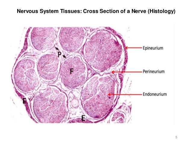
Mammal. Cerebral cortex. Neuron. Silver stain. 125X - Neuron - Mammals - Mammals - Nervous ...
A Labelled Diagram Of Neuron with Detailed Explanations A Labelled Diagram Of Neuron with Detailed Explanations Biology Biology Article Diagram Of Neuron Diagram Of Neuron A neuron is a specialized cell, primarily involved in transmitting information through electrical and chemical signals. They are found in the brain, spinal cord and the peripheral nerves. A neuron is also known as the nerve cell.

worksheet. Nervous System Worksheet. Grass Fedjp Worksheet Study Site
Neuron Diagram & Types | Ask A Biologist Types of Neurons. There are many types of neurons in your body. Each type is specialized to be good at doing different things. Multipolar neurons have one axon and many dendritic branches. These carry signals from the central nervous system to other parts of your body such as your muscles and glands. Unipolar neurons are also known as sensory ...
Activity 7 - Brain & Cranial Nerves
Types of Neurons: Parts, Structure, and Function - Verywell Health Summary. Neurons are responsible for transmitting signals throughout the body, a process that allows us to move and exist in the world around us. Different types of neurons include sensory, motor, and interneurons, as well as structurally-based neurons, which include unipolar, multipolar, bipolar, and pseudo-unipolar neurons.
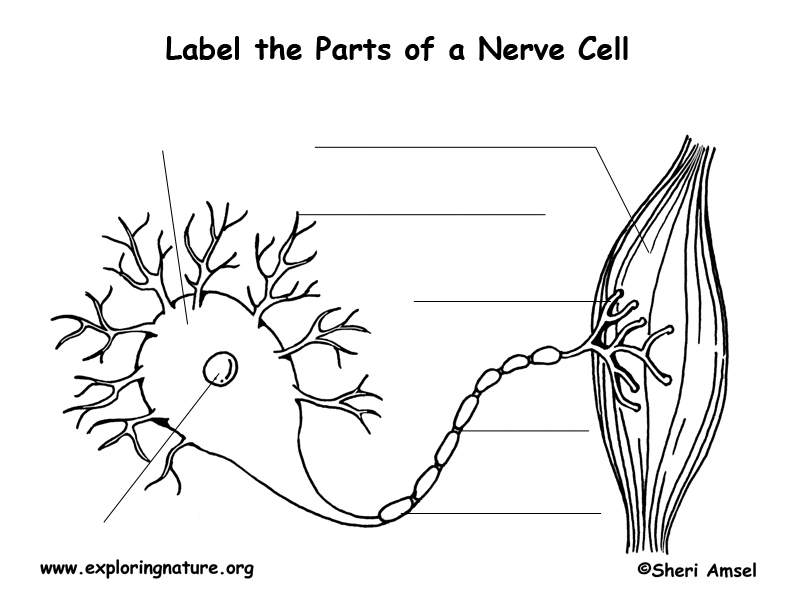
Nerve Cell (Neuron) Labeling Page
Nervous System Anatomy Stock Photos And Images - 123RF Affordable and search from millions of royalty free images, photos and vectors. Photos. Vectors. FOOTAGE. AUDIO. SEE PRICING & PLANS. Support. en ... Nervous system. Human anatomy. Brain, motor neuron, glial and.. Vector. Similar Images . Add to Likebox #85341064 - Neurons cells concept ... BLOOD VESSELS_Labels. Similar Images . Add to Likebox ...

34 Neuron To Label - Labels Design Ideas 2020
A Labelled Diagram of Neuron with Detailed decription A neuron is a type of cell that is largely responsible for conveying information via electrical and chemical impulses. The brain, spinal cord, and peripheral nerves all contain them. The nerve cell is another name for a neuron. The structure of a neuron changes depending on its form and size, as well as its function and location.

2,781 Labeled brain anatomy Images, Stock Photos & Vectors - Shutterstock Labeled brain anatomy royalty-free images. 2,781 labeled brain anatomy stock photos, vectors, and illustrations are available royalty-free. See labeled brain anatomy stock video clips. Set goals and get predicted insights based on performance.
Free Neuron Cliparts, Download Free Neuron Cliparts png images, Free ClipArts on Clipart Library
Wikipedia:Featured picture candidates/Neuron cell Neurons (also known as neurones and nerve cells) are electrically excitable cells in the nervous system that process and transmit information. In vertebrate animals, neurons are the core components of the brain, spinal cord and peripheral nerves. Reason. Bumped into this at COM:FPC. Clear, technically precise and encyclopedic SVG diagram of a ...

Neuron Diagram Unlabeled unlabeled diagram of nerve cell · diagram of neuron without labels. Neuron Anatomy Activity. The parts of the neuron have been labeled. Your challenge is to write the correct name for each part and explain what it does. Dec 27, The diagram below is of a nerve cell or neurone. i. Add the following Neuron. JPG. 2.
Post a Comment for "45 picture of a neuron without labels"