40 fluorescent labels and light microscopy
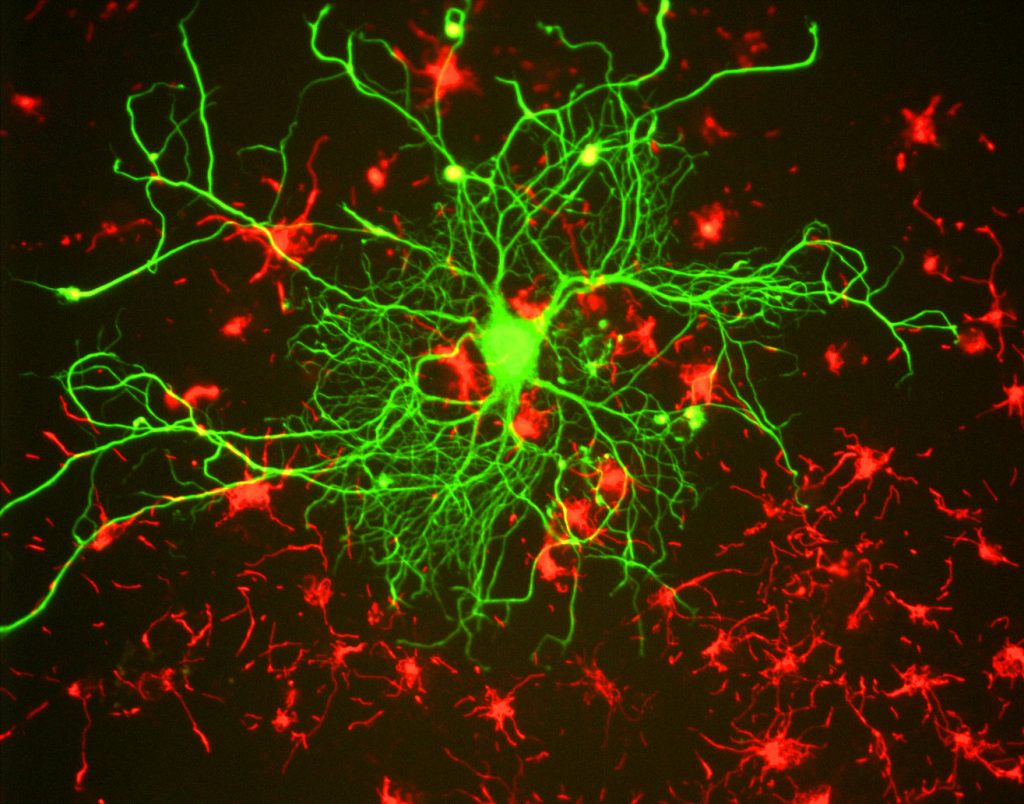
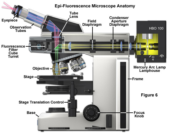
iopscience.iop.org › journal › 2050-6120Methods and Applications in Fluorescence - IOPscience These encompass biological, medical, chemical, material and nano research using experimental, theoretical and data analysis methods, which span probes, spectroscopy, imaging and microscopy. The journal publishes original research articles, topical reviews, tutorials, technical notes and editorial perspectives. biotium.com › technology › cellular-stainsFluorescent Cell Stains for Organelles & Cellular Structures ... “Light-On” LysoView™ 555 “Light-On” LysoView™ 555 is a UV-activatable lysosome stain. In cells, the dye initially shows low fluorescence, but brief exposure to UV excitation from a mercury arc lamp activates bright red fluorescence localizing to lysosomes.
en.wikipedia.org › wiki › Two-photon_excitationTwo-photon excitation microscopy - Wikipedia Two-photon excitation microscopy typically uses near-infrared (NIR) excitation light which can also excite fluorescent dyes. However, for each excitation, two photons of NIR light are absorbed. Using infrared light minimizes scattering in the tissue. Due to the multiphoton absorption, the background signal is strongly suppressed.

Fluorescent labels and light microscopy
› createJoin LiveJournal Password requirements: 6 to 30 characters long; ASCII characters only (characters found on a standard US keyboard); must contain at least 4 different symbols; en.wikipedia.org › wiki › Super-resolution_microscopySuper-resolution microscopy - Wikipedia Integrated correlative light and electron microscopy. Combining a super-resolution microscope with an electron microscope enables the visualization of contextual information, with the labelling provided by fluorescence markers. This overcomes the problem of the black backdrop that the researcher is left with when using only a light microscope. › lifestyleLifestyle | Daily Life | News | The Sydney Morning Herald The latest Lifestyle | Daily Life news, tips, opinion and advice from The Sydney Morning Herald covering life and relationships, beauty, fashion, health & wellbeing
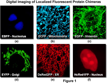
Fluorescent labels and light microscopy. › applications › fretBasics of FRET Microscopy | Nikon’s MicroscopyU The first fluorescent protein biosensor was a calcium indicator named cameleon, constructed by sandwiching the protein calmodulin and the calcium calmodulin-binding domain of myosin light chain kinase (M13 domain) between enhanced blue and green fluorescent proteins (EBFP and EGFP). In the presence of increasing levels of intracellular calcium ... › lifestyleLifestyle | Daily Life | News | The Sydney Morning Herald The latest Lifestyle | Daily Life news, tips, opinion and advice from The Sydney Morning Herald covering life and relationships, beauty, fashion, health & wellbeing en.wikipedia.org › wiki › Super-resolution_microscopySuper-resolution microscopy - Wikipedia Integrated correlative light and electron microscopy. Combining a super-resolution microscope with an electron microscope enables the visualization of contextual information, with the labelling provided by fluorescence markers. This overcomes the problem of the black backdrop that the researcher is left with when using only a light microscope. › createJoin LiveJournal Password requirements: 6 to 30 characters long; ASCII characters only (characters found on a standard US keyboard); must contain at least 4 different symbols;














Post a Comment for "40 fluorescent labels and light microscopy"