42 diagram of the lungs with labels
Heart Diagram with Labels and Detailed Explanation - BYJUS Diagram of Heart. The human heart is the most crucial organ of the human body. It pumps blood from the heart to different parts of the body and back to the heart. The most common heart attack symptoms or warning signs are chest pain, breathlessness, nausea, sweating etc. The diagram of heart is beneficial for Class 10 and 12 and is frequently ... Animal and Plant Cell Worksheets - Super Teacher Worksheets This is a basic illustration of a plant cell with major parts labeled. Labels include nucleus, chloroplast, cytoplasm, membrane, cell wall, and vacuole, and mitochondrion. Use it as a poster in your classroom or have students glue it into their science notebooks.
Circulatory System Diagram - Cardiovascular System and Blood ... How to Make a Circulatory System Diagram. SmartDraw has a number of templates included for circulatory system diagrams, cardiovascular system diagrams, blood circulation diagrams, and more. You don't really have to "draw" them as much as find them and modify them as needed. You can add labels or titles and change the size of symbols as necessary.
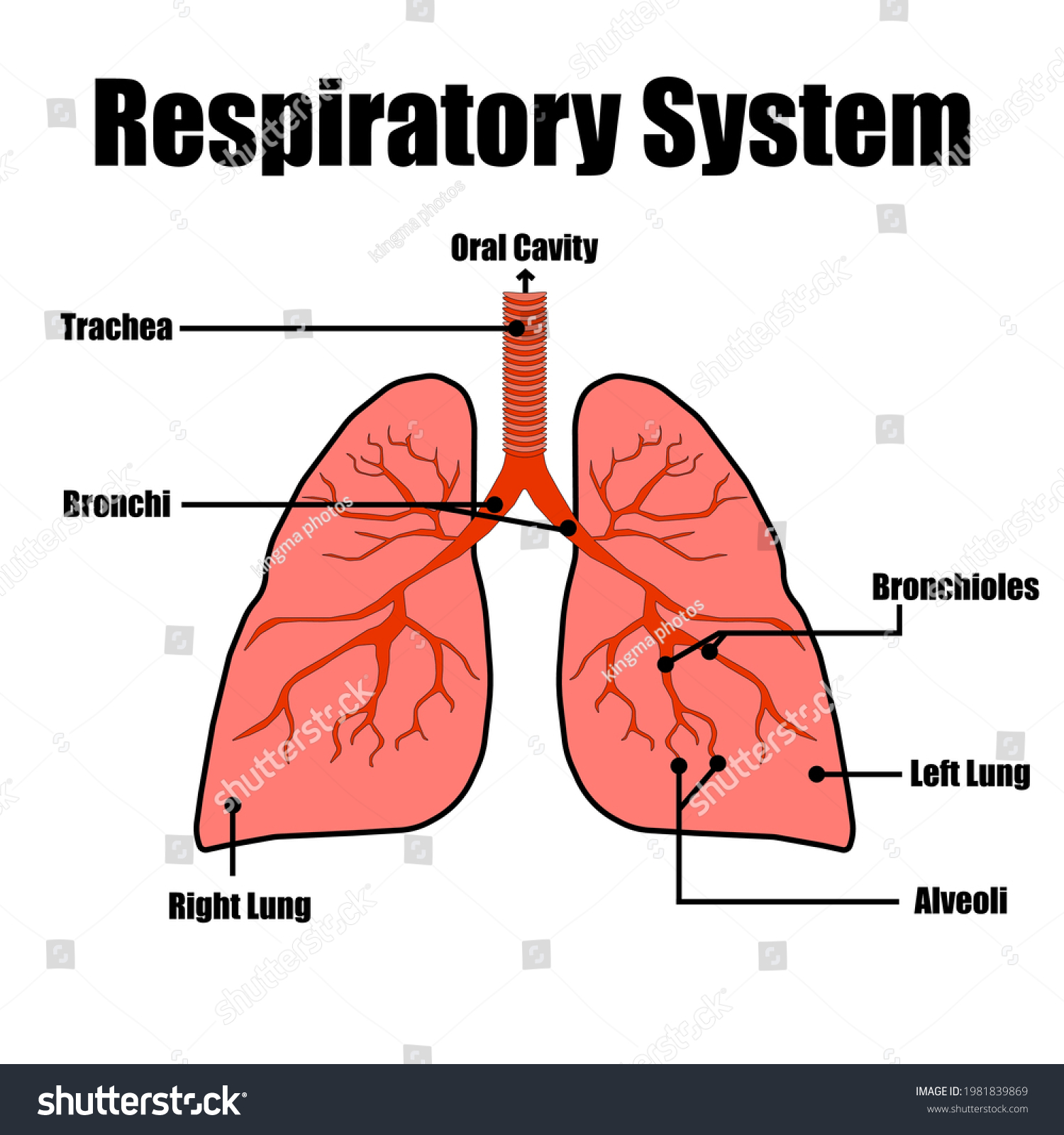
Diagram of the lungs with labels
Diagram Of The Respiratory System With Labels Drawing Illustrations ... A medical diagram showing the lobes of the lungs (organ of the respiratory system) with text labels. Colorful hand drawn illustration of human heart anatomy. Hand drawn illustration of human heart anatomy. Educational diagram showing blood flow with main parts labeled. Vector illustration easy to edit. Fully Labelled Diagram Alveolus Lungs Showing Stock ... - Shutterstock Fully labelled diagram of the alveolus in the lungs showing gaseous exchange. Vector Formats EPS 1114 × 800 pixels • 3.7 × 2.7 in • DPI 300 • JPG Vector Contributor S Steve Cymro Categories: Science , Education Labeled Diagram of the Human Lungs - Bodytomy Given below is a labeled diagram of the human lungs followed by a brief account of the different parts of the lungs and their functions. Each lung is enclosed inside a sac called pleura, which is a double-membrane structure formed by a smooth membrane called serous membrane.
Diagram of the lungs with labels. labeled diagram of the lungs - Microsoft labeled diagram of the lungs Respiratory unlabeled breathing diagrams labeling srinivasa ramanujan hitam 1480 respiratorio anatomie highlands. Respiratory system worksheet. System cardiovascular human heart diagram anatomy blood circulatory flow lungs biology medical coming clipart ventricles allow anatomie body parts diseases Human Heart (Anatomy): Diagram, Function, Chambers, Location ... The left atrium receives oxygenated blood from the lungs and pumps it to the left ventricle. The left ventricle (the strongest chamber) pumps oxygen-rich blood to the rest of the body. The left ... labeled diagram of lungs Lung Anatomy Diagram - Anatomy Diagram Book grekoulas.blogspot.com. lungs. A&P CH22 The Respiratory System Flashcards | Easy Notecards . respiratory system diagram lungs bronchi labeled lung label drag human tree appropriate each bronchiole alveoli where flashcards terminal does division. 5. Frog Dissection - Ms. Spira SNC ... Diagram Of The Respiratory System With Labels Pictures, Images ... - iStock In mammals and most other vertebrates, two lungs are located near the backbone on either side of the heart. Vector graphic. Lungs with Alveoli Labeled CG image of woman's chest area showing both lungs in isolation, with magnified view of alveoli air sacs labeled on faded flesh tone and white.
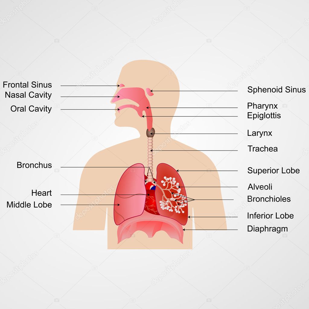
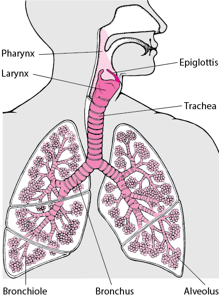
Lung Diagram Labeled | EdrawMax Template in the following lung labeled diagram, we have shown thyroid cartilage, cricoid cartilage, tracheal cartilage, apex, left upper lobe, hilum, left bronchus, oblique fissure, bronchioles, left lower lobe, base of lung, cardiac notch, right lower lobe, oblique fissure, right middle lobe, horizontal fissure, right bronchus, right upper lobe, and … Human Body Worksheets - Easy Teacher Worksheets In the diagram to the left, provide the labels for the structures involved in the reflex act when a person steps on a tack and jerks their leg away. Brain Anatomy Provide the labels for the diagram on the left below and provide descriptions of the functions of each structure on the blank lines. Labeled diagram of the lungs/respiratory system. - SERC View Original Image at Full Size. Labeled diagram of the lungs/respiratory system. Image 37789 is a 1125 by 1408 pixel PNG Uploaded: Jan10 14. Last Modified: 2014-01-10 12:15:34 Anatomy and Physiology 2e - 2e - Open Textbook Library Anatomy and Physiology 2e is developed to meet the scope and sequence for a two-semester human anatomy and physiology course for life science and allied health majors. The book is organized by body systems. The revision focuses on inclusive and equitable instruction and includes new student support. Illustrations have been extensively revised to be clearer and more inclusive. The web-based ...
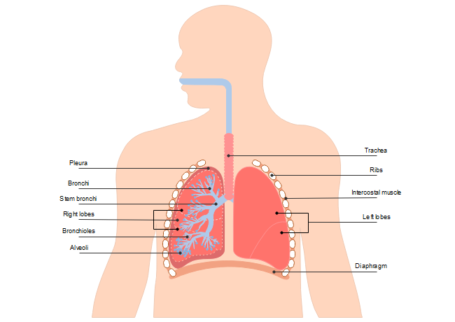
detailed heart diagram labeled Label The Lungs Diagram - Human Anatomy tartrerepub.blogspot.com. respiratory lungs trachea alveoli physiology ribs diaphragm pleural cavity bronchioles libretexts wikieducator larynx bronchi. Coronary Arteries (anterior View). | Download Scientific Diagram . Lung Diagram | Free Lung Diagram Template - Edrawsoft The lung diagram template here clearly presents a pair of spongy on both side of the chest. Simply hitting on the template to learn more parts including pleura, ribs, bronchi, alveoli and more. Feel free to find out more human anatomy templates and symbols in the free download version. Download Template: Get EdrawMax Now! Free Download Diagram Of The Lungs Without Labels Diagram Of The Lungs Without Labels. Respiratory System With Images Respiratory System. Lung Diagram Worksheet With Images Heart Diagram Human Heart. Pin On Nursing School Tips. Respiratory System Diagram. Label The Skeleton With Images Anatomy And Physiology Human. lungs diagram to label respiratory system labels name clipart Posterior view angled to the right hand side of the lungs and ribcage. Trachea respiratory system anatomy bronchi windpipe larynx respiration human parts platypus lungs interactive left function body tube carina superior activity. Free anatomy clipart
Respiratory System 3d Animated Color Labels Stock Vector ...
Lobes of the Lung - SmartDraw Venn Diagram Wireframe Lobes of the Lung Create healthcare diagrams like this example called Lobes of the Lung in minutes with SmartDraw. SmartDraw includes 1000s of professional healthcare and anatomy chart templates that you can modify and make your own. 4/22 EXAMPLES EDIT THIS EXAMPLE Text in this Example: Lobes of the Lung

Anatomy of the Lungs PowerPoint Diagram - PSlides
Lungs diagram - ixwgz.mreds.shop Feb 15, 2022 · Lung Diagram This image details the anatomy of the lung, and specifically highlights the lining of the lungs, known as the pleura, where pleural mesothelioma develops.This is the most common form of the cancer and develops when the mesothelial cells that line the pleura become cancerous and divide uncontrollably.

The Structure Of A Lung With Labeled Parts. Biology Vector ...
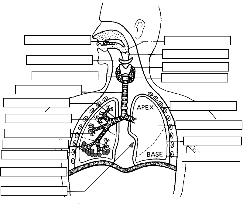
Lung Diagram Labelling Activity | Primary Resources | Twinkl This handy Lung Labelling Worksheet gives your children the opportunity to show how much they've learned about the human lung system. The beautifully hand-drawn illustration shows a lung diagram, labelled with blank spaces where learners can fill in its different components. Encourage your students to work independently and label the parts of the lungs they can see. This teaching resource also ...
Lung Diagram | Free Lung Diagram Template
8,991 Lung Diagram Images, Stock Photos & Vectors | Shutterstock 8,960 lung diagram stock photos, vectors, and illustrations are available royalty-free. See lung diagram stock video clips Image type Orientation Sort by Popular Healthcare and Medical Anatomy Recreation/Fitness Diseases, Viruses, and Disorders lung respiratory system medicine pulmonary alveolus organ human body Next of 90
The Anatomy and Physiology of Animals/Respiratory System ...
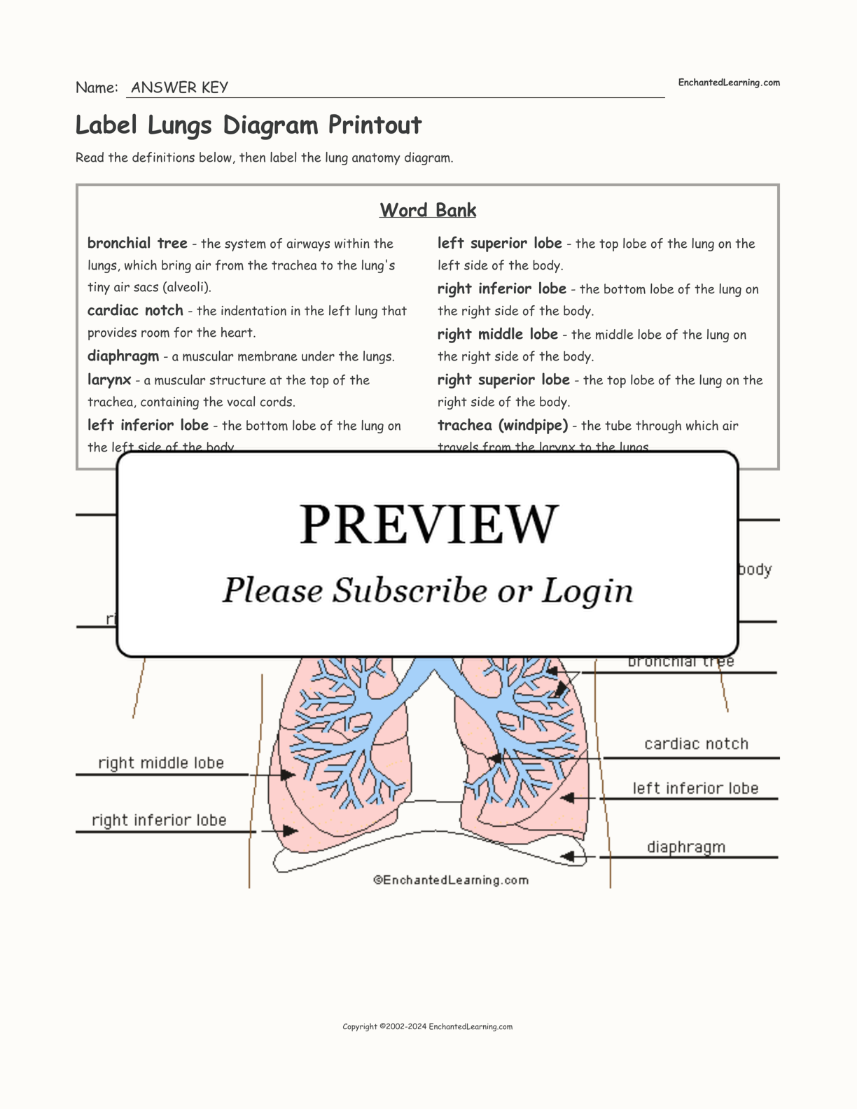
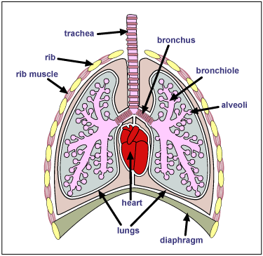
Label Lungs Diagram Printout - Enchanted Learning bronchial tree: the system of airways within the lungs, which bring air from the trachea to the lung's tiny air sacs (alveoli). cardiac notch: the indentation in the left lung that provides room for the heart. diaphragm: a muscular membrane under the lungs. larynx: a muscular structure at the top of the trachea, containing the vocal cords.

lungs diagram | How do we breathe? (Lungs and Pleura ...
labeled diagram of the lungs The Heart Diagrams Labeled and Unlabeled. 7 Pics about The Heart Diagrams Labeled and Unlabeled : Tuberculosis by:kaitlyn nordby, Phenomia Symptoms | HRFnd and also draw a well labelled diagram of the section of an alveolus and the. The Heart Diagrams Labeled And Unlabeled . labeled unlabeled classconnection

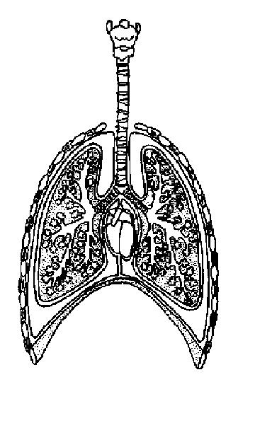
A schematic drawing of the lungs and airway tree in which ...
Diagram Of The Lungs With Labels Labeling Of The Lungs Label The Lungs ... Diagram Of The Lungs With Labels Labeling Of The Lungs Label The Lungs Diagram Diagram Of Lungs With. By admin Apr 15, 2019. Share this page . Post navigation. Lung Lobectomy: What you need to know . By admin. Related Post. Leave a Reply Cancel reply. You must be logged in to post a comment.

Lung Structure | BioNinja
Label the Lungs Diagram | Quizlet Label the Lungs Learn Test Match Created by ohcrawford1 PLUS Terms in this set (9) trachea ... superior lobe of right lung ... middle lobe of right lung ... inferior lobe of right lung ... superior lobe of left lung ... left main (primary) bronchus ... lobar (secondary) bronchus ... segmental (tertiary) bronchus ... inferior lobe of left lung ...
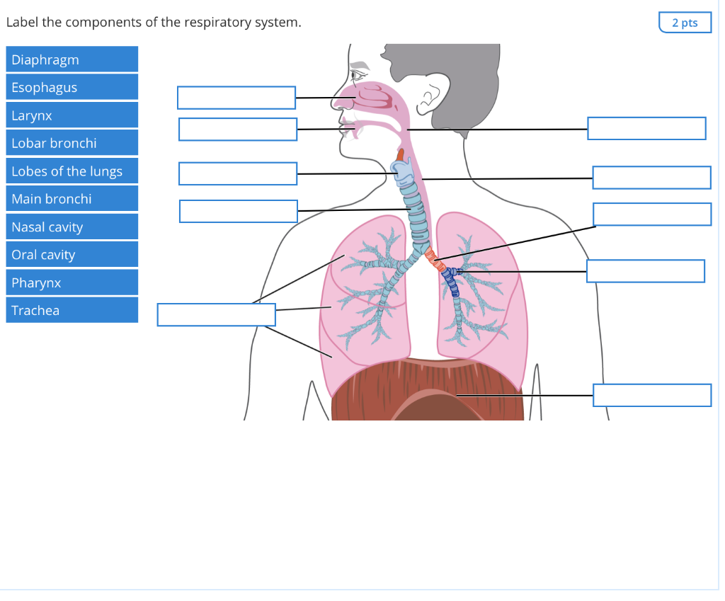
Solved Label the components of the respiratory system. 2 pts ...
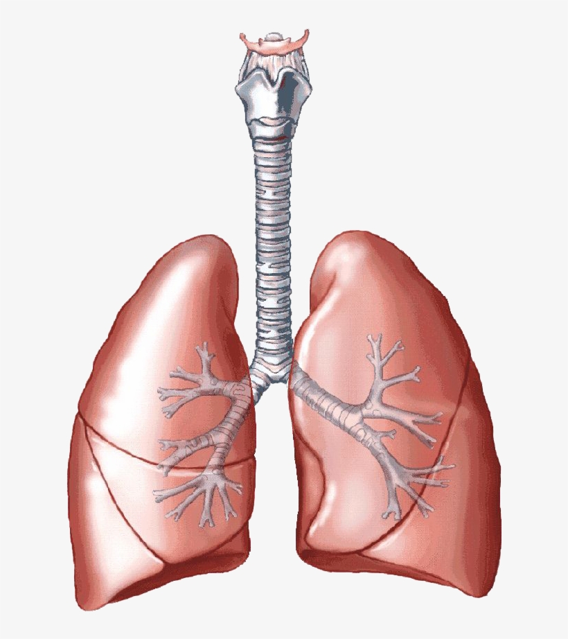
Lung Anatomy, Function, and Diagrams - Healthline The lungs begin at the bottom of your trachea (windpipe). The trachea is a tube that carries the air in and out of your lungs. Each lung has a tube called a bronchus that connects to the...

How to draw and label a lung | step by step tutorial
Diagram of Human Heart and Blood Circulation in It Ventricle contracts and pushes the blood into the pulmonary artery that sends blood to your lungs from where oxygen-rich blood returns to the left ventricle and the process continues. Exterior of the Human Heart A heart diagram labeled will provide plenty of information about the structure of your heart, including the wall of your heart.

File:Respiratory system complete no labels.svg - Wikimedia ...
The Lungs - Position - Structure - TeachMeAnatomy Surfaces. There are three lung surfaces, each corresponding to an area of the thorax. The mediastinal surface of the lung faces the lateral aspect of the middle mediastinum. The lung hilum (where structures enter and leave the lung) is located on this surface.. The base of the lung is formed by the diaphragmatic surface.It rests on the dome of the diaphragm, and has a concave shape.
The lungs | Macmillan Cancer Support
Lungs (Human Anatomy): Picture, Function, Definition, Conditions - WebMD The lungs are a pair of spongy, air-filled organs located on either side of the chest (thorax). The trachea (windpipe) conducts inhaled air into the lungs through its tubular branches, called...
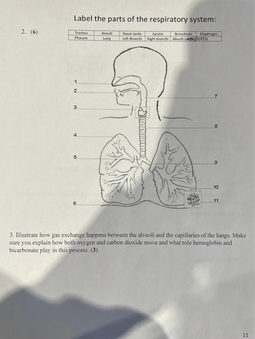
Solved Label the parts of the respiratory system: 2. (6 ...
Sample 1: Heart and Lung Diagram The circulatory and respiratory systems work together to transport oxygen-rich blood through the body. A diagram shows a cross-section of a heart between two lungs. Red arrows show the path of oxygen-rich blood cells. Blue arrows show the path of oxygen-poor blood. Oxygen-rich blood cells travel to the heart from the lungs.

Task 10: Label the chest & lungs (Yr7) Diagram | Quizlet
lungs heart diagram The Heart Diagrams Labeled And Unlabeled . unlabeled labeled diagrams circulatory unlabelled. Sample 1: Heart And Lung Diagram - Accessible Image Sample Book diagramcenter.wpengine.com. lungs heart oxygen diagram lung carbon dioxide through cells epub move sample zoom samplebook wpengine. Rib Cage - Medical Art Library

The Lungs by David Gabb
Lungs Diagram - Human Lungs Anatomy - BYJUS The right lung comprises three lobes - inferior, middle and superior lobe that are distinguished by an oblique and deep horizontal fissure. The left lung has two lobes separated by an oblique fissure. The apexes of the lungs expand above the first rib, while both lungs in the thorax rest with their bases on the diaphragm.

Draw a diagram of human respiratory system and label ...
Diagram Lungs Illustrations & Vectors - Dreamstime Download 2,677 Diagram Lungs Stock Illustrations, Vectors & Clipart for FREE or amazingly low rates! New users enjoy 60% OFF. 195,607,173 stock photos online. ... Labeled diagram with sickness symptoms. Lupus disease vector illustration. Labeled diagram with sickness symptoms, like hair. Different systems of human body diagram. Illustration
Human Lungs Worksheets - Superstar Worksheets
How to Draw a Human Heart: An Easy Step-By-Step Guide - wikiHow Sep 20, 2022 · The heart works like a pump and beats 100,000 times a day. The heart has two sides, separated by an inner wall called the septum. The right side of the heart pumps blood to the lungs to pick up oxygen. The left side of the heart receives the oxygen-rich blood from the lungs and pumps it to the body.

Lung Tissue | BioNinja
How to draw and label a lung | step by step tutorial - YouTube A beautiful drawing of Lung. And it will teach you to draw the lung very easily. Watch the video and please be kind enough to thumbs up my videos. And I will...

Respiratory System Label Me Diagram | Quizlet
Labeled Diagram of the Human Lungs - Bodytomy Given below is a labeled diagram of the human lungs followed by a brief account of the different parts of the lungs and their functions. Each lung is enclosed inside a sac called pleura, which is a double-membrane structure formed by a smooth membrane called serous membrane.

Draw a diagram of human respiratory system and label ...
Fully Labelled Diagram Alveolus Lungs Showing Stock ... - Shutterstock Fully labelled diagram of the alveolus in the lungs showing gaseous exchange. Vector Formats EPS 1114 × 800 pixels • 3.7 × 2.7 in • DPI 300 • JPG Vector Contributor S Steve Cymro Categories: Science , Education

How to draw Lungs diagram | Science drawing, Biology diagrams ...
Diagram Of The Respiratory System With Labels Drawing Illustrations ... A medical diagram showing the lobes of the lungs (organ of the respiratory system) with text labels. Colorful hand drawn illustration of human heart anatomy. Hand drawn illustration of human heart anatomy. Educational diagram showing blood flow with main parts labeled. Vector illustration easy to edit.
Label respiratory system - Teaching resources
Medical illustration of human lungs anatomy with labels ...

Label lungs Diagram | Quizlet

3,561 Respiratory System With Labels Stock Photos, Pictures ...
Label Lungs Diagram Printout - Enchanted Learning

In the diagram below, label the parts of the respiratory ...
IB Biology Notes - 6.4 Gas exchange

draw labelled diagram of lungs - Brainly.in
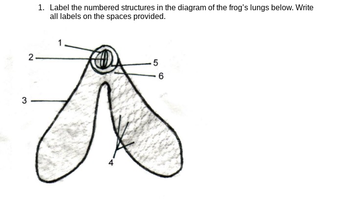
Answered: 1. Label the numbered structures in the… | bartleby
Respiratory System Anatomy - Major Zones & Divisions ...

Draw a diagram of the human respiratory system and label ...

Lesson Worksheet:The Mechanism of Breathing | Nagwa

With the help of labelled diagram explain the structure of ...
Respiratory System Stock Vector Image by ©stockshoppe #9977839
Overview of the Respiratory System - Lung and Airway ...

File:Lungs diagram simple.svg - Wikimedia Commons
Lungs Png Clipart - Anatomy Respiratory System Label Practice ...

Lung diagram | Lungs image | Simple lungs diagram | Biology ...

The Lungs | Anatomy and Physiology II
Anatomy: Respiratory System

Label the Lungs (1) Diagram | Quizlet
Post a Comment for "42 diagram of the lungs with labels"